
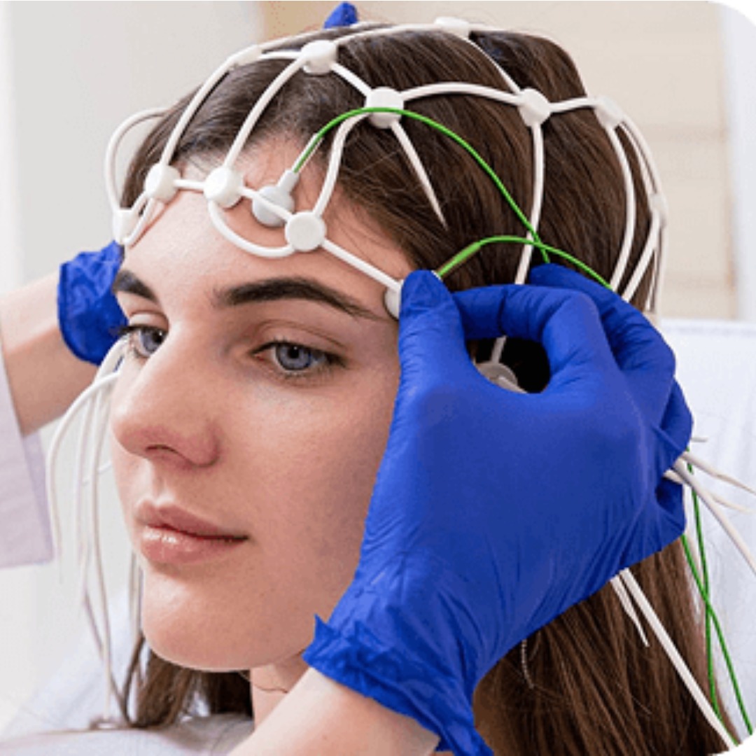
When symptoms such as brain fog, difficulty concentrating, headaches or memory problems continue after a concussion or other health concern, people often want information that goes beyond describing how they feel. qEEG brain mapping is a non-invasive way of recording and analyzing electrical activity produced by the brain.
For Fargo–Moorhead patients considering a qEEG assessment, understanding what the technology measures—and what it cannot determine on its own—is an important first step.
What Does qEEG Mean?
qEEG stands for quantitative electroencephalography. It begins with an EEG recording, which measures the brain’s naturally occurring electrical signals through sensors placed on the scalp. The sensors receive information; they do not send electricity into the brain.
Specialized software then applies mathematical analysis to the recorded EEG data. The results can be displayed as color-coded maps and numerical measurements showing how electrical activity is distributed across different areas and frequency ranges.
This visual display is why qEEG is often called brain mapping. It does not produce an anatomical picture like a CT scan or MRI. Instead, it organizes information about electrical activity recorded during the assessment.
What Happens During a qEEG Assessment?
At Swanson Hyperbarics, qEEG brain mapping in Fargo, ND uses a soft cap fitted with multiple sensors. A trained team member positions the cap on the patient’s head and checks the sensors before recording begins.
The patient then sits quietly while the system records brainwave activity. The recording itself takes approximately 20 minutes. Remaining relaxed and limiting movement are important because blinking, facial tension and body movement can create artifacts that interfere with the data.
Preparation instructions can vary. Patients may be asked to arrive with clean, dry hair and avoid certain hair products. Sleep, caffeine, medications and other factors may influence brain activity, so patients should follow the instructions they receive and provide accurate information. Medication should never be stopped unless the prescribing healthcare provider has specifically advised it.
The procedure is painless, involves no radiation and requires no injections. Once sufficient data have been collected, the software processes the recording for review.
What Does a Brain Map Show?
A qEEG report may display measurements associated with different brainwave frequency ranges and show how activity varies across the recorded areas. Depending on the software and clinical process, results may also be compared with information from a reference database.
These measurements can help a qualified professional identify patterns that may deserve closer attention. They may also create objective baseline data that can be considered alongside symptoms, medical history and other assessments.
However, a colored area on a brain map does not automatically identify a particular disease or reveal the precise cause of a symptom. Similar patterns can occur for different reasons, while sleep, medication, movement or recording quality can affect results. Interpretation requires context and professional judgment.
How Can qEEG Relate to Concussion or Brain-Injury Assessment?
A concussion is a mild traumatic brain injury that can disrupt brain function even when a conventional scan does not show structural damage. CT and MRI remain important when clinicians need to look for bleeding, swelling, fractures or other physical changes, but they measure something different from electrical activity.
qEEG is being researched as a way to study electrical patterns following concussion and traumatic brain injury. It may provide supplementary information during a broader evaluation, particularly when patients report persistent cognitive or neurological symptoms.
Its limitations must also be understood. An American Clinical Neurophysiology Society guideline concluded that current evidence does not support using qEEG by itself to diagnose mild traumatic brain injury or distinguish it reliably from other conditions. qEEG should therefore be viewed as an additional source of information—not as a stand-alone concussion test or a replacement for medical diagnosis.
Why a Broader Evaluation Matters
Brain-related symptoms rarely tell their full story through one measurement. A careful evaluation may consider how the injury occurred, when symptoms began, whether they are changing and how they affect work, sleep, balance, vision, mood and everyday activities.
At Swanson Hyperbarics, qEEG can be considered alongside NeckCare for the patient’s wider clinical information. It examines aspects of visual tracking, reaction time and coordination, while qEEG records electrical brain activity. These services collect different types of information and should be interpreted within the patient’s overall assessment.
Anyone with a recent head injury should first receive appropriate medical evaluation. The CDC advises seeking immediate emergency care for signs such as a worsening headache, repeated vomiting, seizures, weakness, slurred speech, increasing confusion, unequal pupil size or difficulty waking. Brain mapping is not an emergency screening tool.
qEEG Brain Mapping in Fargo, ND
Swanson Hyperbarics provides qEEG brain mapping and NeckCare for patients in Fargo, Moorhead and surrounding communities. The purpose of the assessment process is to gather additional information that may help patients and providers have a more informed conversation about symptoms, function and possible next steps.
Before scheduling, patients should ask what the assessment includes, who will interpret the results and how the findings will be used. Clear expectations are especially important because qEEG is an adjunctive tool rather than an independent medical diagnosis.
To learn whether qEEG may be appropriate for your situation, contact Swanson Hyperbarics to schedule a consultation.
Summary
qEEG brain mapping is a non-invasive process that records electrical activity from the scalp and converts the information into measurable patterns and visual maps. It may contribute useful data to a broader brain-health or brain-injury assessment, but it cannot independently diagnose concussion, TBI or another neurological condition. The most responsible use of qEEG combines careful recording, qualified interpretation, medical history and other relevant clinical findings.